Honing in on the Biological Markers of Autism

Back To Top
Honing in on the Biological Markers of AutismHoning in on the Biological Markers of Autism

The prevalence of autism spectrum disorders, or ASD, is staggering — an estimated 1 out of 88 children have some form of ASD. As the word “spectrum” in the name suggests, ASD varies in its range and severity among those affected. The various forms of the disorder share some common characteristics, however, one of which centers on language and communication deficits.

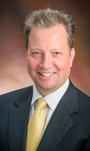
A Children’s Hospital investigator is taking a unique approach to determine how those with autism process sounds, words, and images, and then use those findings to develop potential interventions. Rather than looking at autism from a behavioral or clinical perspective, Timothy Roberts, PhD, vice chair of the Department of Radiology, is using sophisticated imaging to hone in on the biological basis for autism.

Since 2007, Dr. Roberts, who also holds the Oberkircher Family Endowed Chair in Pediatric Radiology, has used magnetoencephalography (MEG) and advanced magnetic resonance imaging (MRI) to look at the “signatures” of brain functioning to reveal the biological nature of ASD. Dr. Roberts and his team observe not just where in the brain things are happening but also the differences in the timing, or frequency, of these signals. These split-second differences can lead to auditory processing delays seen in some children with ASD.

But knowing some of the autism signatures is not enough to move on to possible interventions. What’s needed, Dr. Roberts says, is “a little bit of biology” that can shed light on what’s giving rise to those signatures in the first place. Identifying and understanding potential biomakers may then lead to more options for treating ASD.

With a multi-modal approach using MEG and MRI, Dr. Roberts and colleague James (Chris) Edgar, PhD, have found a biomarker in the white matter of the brain that may be the cause of delayed auditory processing. And he’s found a second one in a neurotransmitter called GABA. An inhibitory neuron, GABA must be in balance with another neurotransmitter — the excitatory glutamate.

“Those with ASD who have lower levels of GABA end up with a more ‘noisy brain,’” Dr. Roberts says. “This is a problem because it means these children are trying to encode their perceptual reality, their world — and they are trying to encode it with rhythms in a sea of noise.”

The tremendous variability, or heterogeneity, seen in children with ASD suggests that the problem for some may arise from abnormal white matter development and for others may stem from reduced levels of GABA. Knowing the biological differences at play has led Dr. Roberts toward existing drugs that target GABA — the effectiveness of which he could measure using advanced imaging techniques — and toward developing a novel intervention for those whose ASD may arise from auditory processing difficulties.

“Five years ago we didn’t have signatures,” says Dr. Roberts. “Now we not only have signatures but we’ve transitioned into candidates for biomarkers. And we’ve filed a provisional patent application on the use of MEG as a biomarker for clinical trial design and, ultimately, therapeutic monitoring for those with ASD who have a GABA-glutamate imbalance.”

For those whose ASD stems from an abnormal structural connectivity in the language pathway of the brain, Dr. Roberts and his colleagues, including David Embick, PhD, from the Department of Linguistics at the University of Pennsylvania, are looking at ways to enhance what is called “repetition priming.” It works like this: saying the word “cat” likely conjures an image of a four-legged, green-eyed, furry animal with pointy ears and whiskers. The specifics of the cat’s characteristics — it’s color and so forth — vary among people, but the overall representation of a cat is the same for all of us.

For children with more severe forms of ASD whose problems arise from the brain’s language pathway, significant amounts of time and energy may be spent focusing on the details and differences in how people actually say the word “cat” — our individual intonations and so forth — and as a result they create separate representations each time they hear the same word.

“These children may be working too hard on the details and are unable to form an object in their mind or categorize it,” Dr. Roberts says. “They may be unable to group things together or cluster them and therefore are treating them all as different. It just becomes a multi-sensory overload.”

Repetition priming speeds up our responses to things that are similar. If children with ASD aren’t able to grow a healthy language because they are doing too much work processing the myriad stimuli they receive, then helping them “cluster” these representations would help them make advance their language and communication.

Drs. Roberts and Embick are developing a device along the lines of a hearing aid (also with a provisional patent application filed) that would filter the incoming language, remove the subtle variations in how we all speak (therefore making everyone sounds the same) and produce one voice that would help children with ASD better form the representations that build their language and vocabulary. And by watching key parts of the brain through imaging, Dr. Roberts can measure whether it’s working.

“What’s great about this multi-modality approach is that we are harnessing cutting-edge imaging technology and using what we learn to forge a path that can make a real difference in the lives of children with autism spectrum disorders,” says Dr. Roberts. “It’s an exciting and promising time.”

Treating the Chicken with the Egg

Back To Top
Treating the Chicken with the EggTreating the Chicken with the Egg

The term “stem cell,” stammzellen, was first used in 1868 by the German biologist Ernst Haeckel to describe the original, unicellular progenitor from which Dr. Haekel supposed all multicellular plant and animal life might have descended. The question, Dr. Haeckel asked, was where that progenitor — the original stem cell — came from in the first place. The chicken or the egg?

Since then, just what defines a stem cell has undergone a few changes. The evolutionary sense of Dr. Haeckel’s term has been dropped, but the sense of stem cells being precursor cells, able to become specialized through the process known as differentiation, remains. Because of their ability to become many types of cells and to renew themselves, stem cells hold enormous promise in understanding and treating a variety of diseases.

What’s more, researchers have identified several different types of stem cells. These include what is perhaps the most popularly known type of stem cell, embryonic stem cells (ESCs), which as their name suggests are derived from embryos. Most often, these come from embryos that have been fertilized through in vitro fertilization and then donated for research purposes.

Another type of stem cell, somatic stem cells, are rare, undifferentiated cells found among other differentiated cells. Also called adult stem cells, there are several types of somatic stem cells: hematopoietic stem cells can differentiate into every type of blood cell, while mesenchymal stem cells can become fat, cartilage, and bone cells.

But in the past decade, researchers have detailed another type of stem cell: induced pluripotent stem cells, or iPSCs. Differentiated adult cells that have been “reprogrammed” and forced to express genes, these cells are capable of developing into many or even all cell types. During fiscal 2013, The Children’s Hospital of Philadelphia’s Mitchell J. Weiss, MD, PhD, published two studies of using iPSCs to study the rare congenital blood disorder Diamond Blackfan Anemia and the childhood cancer juvenile myelomonocytic leukemia.

In the anemia study, Dr. Weiss and his colleagues — including investigators Monica Bessler, MD, PhD, and Philip J. Mason, PhD — removed fibroblasts from Diamond Blackfan Anemia patients and reprogrammed the cells into iPSCs. As those iPSCs were stimulated to form blood tissues, like the patient’s original mutated cells they were deficient in producing red blood cells. However, when the researchers corrected the genetic defect that causes the disorder, the iPSCs developed into red blood cells in normal quantities.

“The technology for generating these cells has been moving very quickly,” said Dr. Weiss. “These investigations can allow us to better understand at a molecular level how blood cells go wrong in individual patients — and to test and generate innovative treatments for the patients’ diseases,” he added.

And in April of 2012, Paul J. Gadue, PhD, published a study detailing a brand new type of stem cell, which the investigators call endodermal progenitor (EP) cells. Produced from ESCs and iPSCs, EP cells have two advantages over these other stem cell types: they do not form tumors when transplanted into animals, and they can form functional pancreatic beta cells in the laboratory. Both ESCs and iPSCs in the undifferentiated state will form a type of tumor called a teratoma when transplanted in animal studies, so it has been critical that any cell generated from ESCs or iPSCs and used for transplantation is purified to exclude undifferentiated cells with tumor-forming potential, Dr. Gadue pointed out.

In addition to producing beta cells, the researchers also directed EP cells to develop into liver cells and intestinal cells — both of which normally develop from the endoderm tissue layer early in human development. The challenge, Dr. Gadue said, has been to differentiate stem cells into one particular cell type in culture, as ESCs and iPSCs can form any cell type in the body. EP cells seem to be limited to cells of the endodermal lineage such as liver, pancreas, and intestine, making it easier to generate pure populations of cells from these organs.

“Our cell line offers a powerful new tool for modeling how many human diseases develop,” said Dr. Gadue, who along with Deborah L. French, PhD, is the co-director of CHOP’s human embryonic stem cell/induced pluripotent Stem Cell (hESC/iPSC) core facility. “Additionally, pancreatic beta cells generated from EP cells display better functional ability in the laboratory than beta cells derived from other stem cell populations.”

In a follow-up review published in fiscal 2013 in Current Opinion in Cell Biology, Dr. Gadue and colleagues discussed the generation of endodermal cells from pluripotent stem cells, including EP cells. Stem cells such as EP cells “can be expanded robustly in culture” and “provide a powerful system to study and model human diseases in vitro, as well as generating a source of cells for transplantation.”

Indeed, there is a big push to use stem cells to perform disease modeling with human cells as opposed to mice, as well as to use stem cells to perform drug and toxicity screenings, Dr. Gadue pointed out, noting that while mouse models offer an incredibly valuable resource they are not perfect and using human cells when possible is important. And once they are created, EP cells can be expanded almost without limit — to the point that Dr. Gadue estimates his laboratory has grown “trillions” of cells — which offers researchers a wealth of research resources.


Going forward, Dr. Gadue, has been working with a number of colleagues, including endocrinologist Diva D. De León-Crutchlow, MD, to better understand and hopefully develop treatments for diabetes.

Collaboration Propels Innovation, Novel Drug Development

Back To Top
Collaboration Propels Innovation, Novel Drug DevelopmentCollaboration Propels Innovation, Novel Drug Development

Numerous research teams at Children’s Hospital work tirelessly year in and year out to uncover the inner workings of biological systems and the causes behind diseases. Armed with their discoveries, the next step may involve using that knowledge to develop new therapeutics to bring to a patient’s bedside.

It sounds like a simple and straightforward approach but, in reality, it isn’t.

Navigating the complex landscape of drug development to treat diseases is a long and detailed process that brings no guarantee of success. Resources to fund the development needed to bridge the gap between the discovery phase and clinical trials are often not readily available. Close collaborations with the pharmaceutical industry are helping to speed the pace of innovation and drug development.

As an example, Children’s Hospital and Pfizer, Inc. joined forces in FY13 to translate biomedical discoveries into novel treatments. The Hospital joined the Centers for Therapeutic Innovation (CTI) network, a novel collaboration model built by Pfizer that brings academic researchers together with Pfizer scientists to expedite the pace of innovation.

“We are excited to have this opportunity to accelerate the process of moving scientific insights toward therapies that healthcare providers can offer in the clinic,” said Philip R. Johnson, MD, chief scientific officer and executive vice president at Children’s Hospital. Dr. Johnson is one of CHOP’s representatives on a joint steering committee with Pfizer representatives that will direct CTI’s activities in Philadelphia.

This expedited timetable that is part of the CTI network is much faster than the typical schedule for federally sponsored research. Many partnerships between private industry and academia that focus on one highly defined end-product; however, CTI is designed to identify cutting-edge areas of research in areas of high unmet need that hold strong potential for therapeutic interventions. The goal is to advance a project into a Phase 1 clinical trial.

Children’s Hospital is only the second pediatric center to participate in the CTI network, which has established partnerships with 21 academic medical centers throughout the United States.

“Working with leading academic researchers is a key part of the CTI model,” said Anthony Coyle, PhD, CTI’s Chief Scientific Officer. “CHOP’s world-class reputation as a leading research hospital means it is an ideal partner for CTI as we continue our determined efforts to translate exciting science into effective medicines for patients.”

Tech Transfer By the Numbers

Back To Top
Tech Transfer By the NumbersTech Transfer By the Numbers

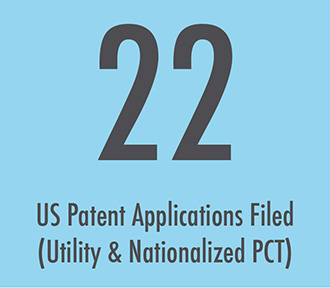
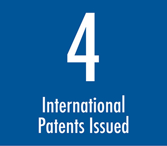
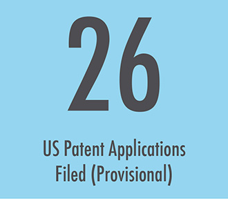
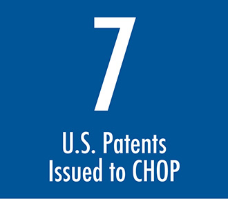
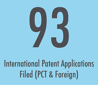
Stats

  • stats1
  • stats2
  • stats3
  • stats4
  • stats5
  • stats6

List of Patents

U.S. Patent No. 8,236,764
Rodney Camire, PhD, and Valder Arruda, MD, PhD

This patent describes methods for creating stable activated coagulation Factor V for treatment of hemophilia A or B. CHOP researchers have devised several potential treatments for hemophilia and this patent covers one such potential treatment.

U.S. Patent No. 8,252,281
Richard Levy, MD, and Clifford Deutschman, MD, MS, FCCM

Drs. Levy and Deustchman have devised a method for treatment of cardiac dysfunction caused by sepsis and associated conditions such as Sepsis-associated Inflammatory Syndrome (SIRS) and Multiple Organ Dysfunction Syndrome (MODS). The treatment entails administration of cytochrome C, a mitochondrial protein involved in cellular respiration, either alone or in combination with ascorbate. This method can also be a potential treatment for cardiac dysfunction associated with trauma, ischemia, inborn errors of metabolism, cardiomyopathies, et cetera.

U.S. Patent No. 8,334,275
Terry Finkel, MD, PhD, and Jiyi Yin

This patent expands previously obtained protection for a method of inducing apoptosis, otherwise known as programmed cell death, in cells infected with HIV as a potential treatment for the disorder.

U.S. Patent No. 8,329,168
Sriram Krishnaswamy, PhD, Elsa Bianchini, PhD, and Steven Orcutt

Proteins of the coagulation pathway are inefficiently converted to the active forms and often require co-factors to do so. This invention related to modification of a variant form of the thrombin zymogen/protease complex to facilitate production of the active polypeptide as a potential treatment for hemophilia.

U.S. Patent No. 8,383,386
Rodney Camire, PhD

This represents another treatment for hemophilia as well as an antidote for over-administration of anti-coagulants. The inventors have created a novel variant of Factor Xa, a component of the coagulation pathway. This invention is licensed and a phase I clinical trial may begin in during FY14.

U.S. Patent No. 8,263,127
Ivan Alferiev, PhD, Ilia Fishbein, MD, PhD, Michael Chorny, PhD, Robert Levy, MD, Benjamin Yellen, PhD, and Darryl Williams, PhD

There is an urgent need for better interfaces between implantable medical devices and tissues. A common cause of implantable device failure relates to inflammation of the surrounding tissues. This patent describes a photo-activatable bio-polymer that could be used to coat the devices and reduce the likelihood of failure.

U.S. Patent No. 8,461,125
Michael Grunstein, MD, PhD

Alternatives to use of corticosteroid treatment of asthma are badly needed. These treatments can have serious side effects and are not always efficacious. This patent describes novel siRNA duplexes directed against CD-23 that will inhibit IgE binding to airway smooth muscle, thereby ameliorating the airway’s response to inhaled allergens.